For every 1,000 elective abdominal cases, somewhere between 4 and 12 patients will experience fascial wound separation, a rate of 0.4% to 1.2%. Emergency laparotomies push that number to 12%.
Those numbers make fascial closure an essential part of every abdominal procedure. The technique and suture material a surgeon chooses directly influence whether the fascia holds or fails.
This post covers fascial closure techniques, suture selection, port-site closure thresholds, and how skin closure after fascial repair affects overall outcomes.
Key Takeaways
- Hernia rates dropped from 21% to 13% when the STITCH trial switched from large-bite to small-bite closure, and a 2025 meta-analysis of 41 RCTs confirmed the same benefit across a broader patient population.
- Slowly absorbable sutures alone don't outperform fast-absorbable sutures for hernia prevention; the benefit only appears when paired with the small-bites technique.
- Nearly all port-site hernias develop at trocar sites of 10 mm or larger, and current guidelines call for fascial closure at every port that size and above.
- Among all modifiable risk factors for incisional hernia, surgical site infection has the greatest impact, according to hernia society guidelines.
- Skin closure with an absorbable subcutaneous stapler averaged 16.2 minutes across over 4,300 cases, while hand-sewn subcuticular suture took 36.5 minutes, with no meaningful difference in infection rates.
What Is Fascial Closure?
Fascial closure is the surgical approximation of the fascia, the strong fibrous layer beneath the skin and fat that holds the abdominal wall together. After any procedure that opens the abdomen, this layer must be closed under tension to restore structural integrity and prevent hernia formation.
The fascia bears the greatest mechanical load of any closure layer. When it fails, surgeons face either acute wound separation or incisional hernia developing over the months that follow. Between 0.4% and 1.2% of elective abdominal patients experience wound separation, and that number jumps to 12% in emergency cases. Mortality after fascial failure exceeds 21%.
Long-term hernia formation is just as concerning. Out of 14,618 patients tracked across 56 publications, 12.8% developed an incisional hernia by roughly two years after midline laparotomy. Patients with elevated BMI or aortic aneurysm history face even steeper odds, with hernia rates approaching 40%.
What Are the Best Suture Techniques for Fascial Closure?
Small-bites closure with a slowly absorbable monofilament suture is the current evidence-based standard for elective midline laparotomy. The European and American Hernia Societies issued a joint guideline in 2022 endorsing this approach. It calls for bites of 5 to 9 mm from the wound edge, spaced 5 mm apart, grabbing aponeurosis only.
Small-Bites vs. Large-Bites Technique
The STITCH trial made the case for small-bite closure. Among 560 patients randomized across multiple centers, 13% of those in the small-bites arm developed an incisional hernia at one year. In the large-bites arm, that figure was 21%. The trade-off was more stitches per closure (a mean of 45 vs. 25) and a few extra minutes on the fascia (14 vs. 10 minutes), but the suture-to-wound ratio improved from 4.3 to 5.0.
Those results held up a decade later. A 2025 meta-analysis pooling 41 RCTs and 9 prospective cohort studies across 2,360 patients found the same pattern. Small-bites closure with a slowly absorbable suture produced lower hernia rates, fewer combined wound separation and hernia events, and reduced surgical site infection compared with large-bites closure.
Continuous vs. Interrupted Closure
Whether a surgeon runs a continuous stitch or places interrupted sutures doesn't appear to change outcomes. Across 14 studies and 5,939 patients in the same 2025 meta-analysis, hernia and wound separation rates were comparable between the two patterns. In practice, continuous closure is faster and uses less suture material. Either pattern works when the bite size and suture-to-wound ratio are correct.
The practical takeaway is straightforward. Bite size and suture-to-wound ratio drive outcomes more than the choice between continuous and interrupted patterns. Surgeons who adopt small-bite closure can use whichever pattern fits their workflow, as long as they maintain the recommended 5 mm spacing and 5 to 9 mm bite depth.
Which Suture Materials Work Best for Fascial Closure?
Slowly absorbable monofilament sutures, such as polydioxanone (PDS) and polyglyconate (Maxon), are the recommended choice for fascial closure in elective midline laparotomy.
One important nuance from a 2025 meta-analysis is that switching to a slowly absorbable suture, by itself, didn't reduce hernia rates. Across 1,177 patients in 5 studies, slowly absorbable materials performed no better than fast-absorbable ones when the bite technique stayed the same. The benefit only showed up when surgeons paired the slowly absorbable suture with small-bite closure. The suture material and the closure technique work together, and changing one without the other doesn't yield the same result.
Monofilament sutures also carry a lower infection risk than braided alternatives. Braided sutures have a larger surface area that can harbor bacteria, making monofilament the preferred choice for abdominal wound closure where surgical site infection is a concern. For surgeons comparing non-absorbable suture options, the current evidence favors slowly absorbable materials for routine fascial closure rather than permanent sutures.
How Does Fascial Closure Differ in Laparoscopic vs. Open Surgery?
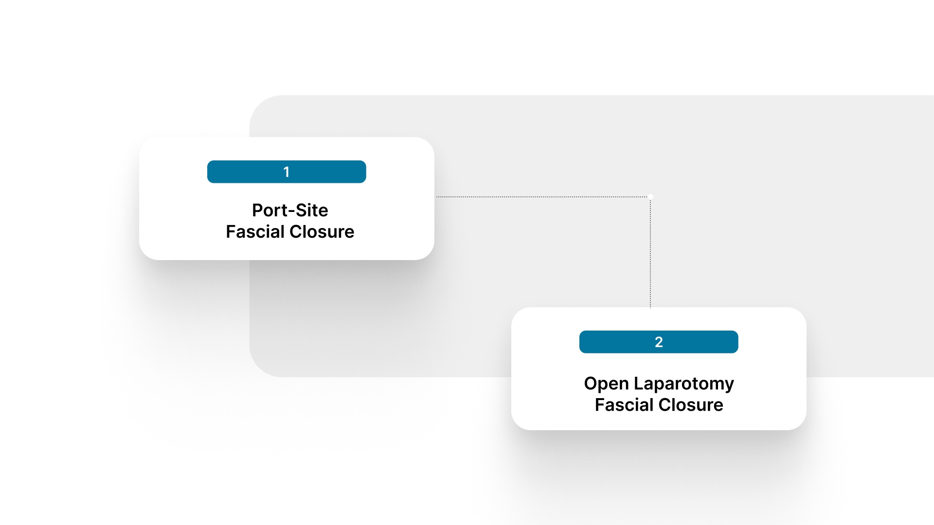
The same principles apply to both settings, but the access constraints and hernia risk profile differ. Open laparotomy involves a long midline incision where the small-bites technique is applied directly under full visualization. Laparoscopic procedures create smaller fascial defects at trocar sites, and the decision to close them depends on port size and location.
Port-Site Fascial Closure
Port-site hernias are relatively uncommon overall, developing in 0.5% to 2% of laparoscopic patients according to a systematic review of over 31,000 cases. Nearly all of them (96%) develop at ports of 10 mm or larger, and the umbilical port is the single most frequent site at 82%.
Current EHS/AHS guidelines are clear on the threshold. Every trocar site of 10 mm and above should get a fascial closure, particularly after single-incision laparoscopic surgery and at the umbilicus. Surgeons can close using a suture-passer device or place sutures directly under visualization.
Open Laparotomy Fascial Closure
Midline laparotomy closure follows the small-bites protocol outlined in the preceding sections. The key technical points remain consistent, including slowly absorbable monofilament suture, 5 to 9 mm bites incorporating aponeurosis only, stitches placed 5 mm apart, and a suture-to-wound length ratio of at least 4:1. For surgeons who use a fascial closure device to assist with port-site or laparotomy closure, the same bite-size and spacing principles apply regardless of the instrument used.
What Are the Risk Factors for Wound Separation After Fascial Closure?
Wound separation after fascial closure results from a combination of patient-level and technical factors. Identifying modifiable risks before surgery and optimizing closure technique together reduces the likelihood of failure.
Patient-level risk factors for increased wound separation include the following.

- Preoperative albumin below 3 g/dL: Low albumin is associated with impaired wound healing and higher rates of fascial wound separation.
- Obesity: Higher BMI increases mechanical tension on the closure and is consistently linked to elevated hernia rates. In high-risk populations, incisional hernia rates can reach approximately 40%.
- Emergency surgery: Wound separation rates in emergency laparotomies are significantly higher than in elective cases, reflecting both patient acuity and suboptimal closure conditions.
- Intraoperative drain placement: Drains placed through or near the fascial closure can weaken the repair and increase infection risk at the closure site.
On the technical side, suture material choice, bite size, and closure tension all influence outcomes. Of all the modifiable risk factors for incisional hernia, surgical site infection has the single biggest impact according to the 2022 EHS/AHS guideline. Choosing a monofilament suture, maintaining proper skin preparation, and following antibiotic prophylaxis protocols all directly protect the fascial closure.
The guideline recommends that surgeons assess and address modifiable risk factors before elective surgery whenever possible. Nutritional optimization, smoking cessation, and glycemic control all contribute to a fascial closure that holds.
How Does Skin Closure After Fascial Repair Affect Outcomes?
Once the fascia is secured, the skin layer still needs closure. The method used here affects OR time, scarring, and postoperative follow-up burden.
Traditional metal staples close skin quickly, but they require a return visit for removal within 7 to 10 days and leave characteristic railroad-track scars at each puncture site. Hand-sewn subcuticular suture avoids surface marks but adds meaningful closure time, particularly on longer incisions.
Bioabsorbable subcutaneous skin closure offers a different approach. Fasteners are placed under the skin, dissolve naturally, and require no removal visit. Across 4,311 mastectomy cases, skin closure with an absorbable stapler took an average of 16.2 minutes. Hand-sewn subcuticular suture on the same type of incision averaged 36.5 minutes, roughly 20 minutes longer per case. Infection rates were virtually identical between the two groups (0.38% vs. 0.36%).
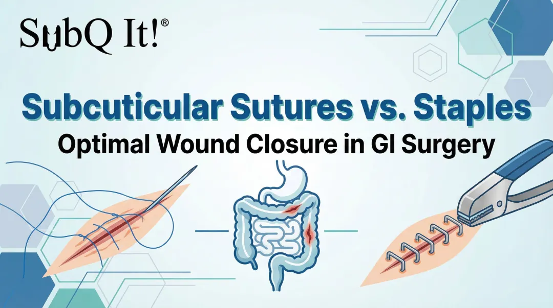
SubQ It! is a bioabsorbable wound closure system that places dissolving dermal fasteners subcutaneously (under the skin) to close incisions. The SubQ It! SU-10 handles incisions up to 10 cm for laparoscopic port sites and smaller incisions, while the SU-25 closes incisions up to 25 cm for longer open procedures. For surgeons performing sutureless skin closure after fascial repair, this approach removes the return visit, reduces surface scarring, and recaptures OR time on the skin layer.
Can Faster Skin Closure Reduce OR Costs After Fascial Repair?
OR time carries a real cost. Every additional minute spent on closure adds to facility expenses and limits how many cases a surgeon can complete in a day. Those costs are especially visible in private practices and ambulatory surgical centers, where case throughput directly affects revenue.
When an absorbable subcutaneous stapler cuts skin closure time by roughly 20 minutes compared to hand-sewn suture, the savings add up across a full operative schedule. For private-practice surgeons and ambulatory surgical centers, that recovered time can translate into additional case capacity over the course of a day or week.
The economic value extends beyond the OR itself. Removing the staple-removal visit from the postoperative workflow saves staff time, frees clinic appointment slots, and reduces the logistical burden on patients. For surgeons looking at the full cost picture of abdominal closure, the skin layer is one area where a disposable skin stapler designed for subcutaneous placement can improve efficiency without compromising wound integrity.
Final Thoughts
Fascial closure is one of the most studied steps in abdominal surgery, and the evidence has converged on a clear recommendation. Small-bites technique with a slowly absorbable monofilament suture, maintaining a suture-to-wound ratio of at least 4:1, is the standard. Applying that consistently, whether at a midline laparotomy or a 10 mm trocar site, is the most reliable way to reduce wound separation and incisional hernia.
The closure doesn't end at the fascia. Skin closure choices affect OR time, scarring, and postoperative follow-up. Surgeons who invest in evidence-based fascial technique can apply the same thinking to the layer above, choosing a closure method that matches the quality of the repair underneath.
SubQ It! is a bioabsorbable wound closure system that places dissolving fasteners under the skin, requiring no removal visit and leaving no railroad-track scars. It works across incision sizes, from laparoscopic port sites (SU-10, up to 10 cm) to longer open incisions (SU-25, up to 25 cm). Contact us today to see if it's the right fit for your practice.
FAQs
1. Can You Use Absorbable Sutures for Fascial Closure?
Yes, slowly absorbable monofilament sutures are the current standard for elective midline fascial closure. The key is pairing them with the small-bites technique, since the suture material alone doesn't drive outcomes without the correct bite pattern.
2. Should Surgeons Use Monofilament or Braided Suture for Fascia?
Monofilament is generally preferred because it has a smoother surface that harbors fewer bacteria. Braided sutures offer good handling characteristics, but the higher infection risk makes monofilament the safer default for fascial closure.
3. Does Fascial Closure Technique Change for Repeat Laparotomies?
Yes, repeat laparotomies often involve scarred or weakened fascia that requires careful identification of healthy tissue edges. The same small-bites principles apply, but surgeons may need wider margins to reach tissue with adequate tensile strength.
4. What Causes Early Wound Separation After Surgery?
Early wound separation is typically caused by technical factors such as inadequate bite depth, excessive closure tension, or use of a suture material that loses strength too quickly. Patient factors like malnutrition, uncontrolled diabetes, and severe coughing also contribute.
5. How Do You Close Fascia in Obese Patients?
Obese patients benefit from the same small-bite, slowly absorbable monofilament approach used in standard-risk patients. Ensuring adequate visualization of the fascial edges and avoiding incorporation of subcutaneous fat into the closure are particularly important in this population.
6. Does the Type of Skin Closure Affect Fascial Healing?
No, skin closure method doesn't directly influence fascial healing. It does affect total OR time, surface scarring, and whether the patient needs a return visit for staple removal, all of which matter for overall surgical efficiency and patient satisfaction.